ACCESSORIES
April 4, 2024
beurer – BM28
April 8, 2024
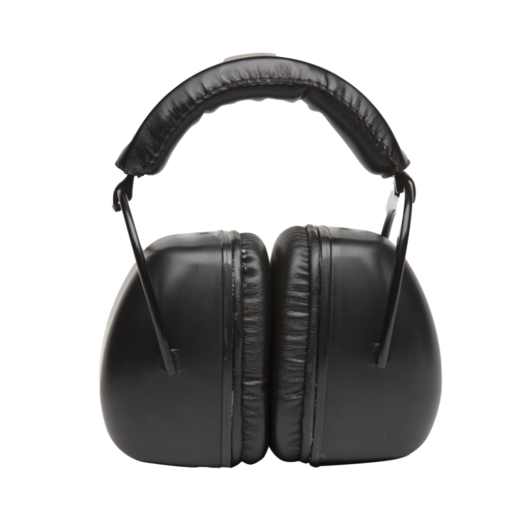
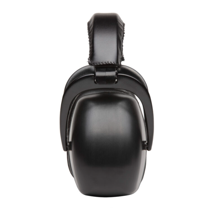
Pro Ears MRI Safe Ultra 30 MRI Kit
ANSI specification S3.19-1974 requires patients undergoing MRI to be provided with noise reduction devices offering a minimum of 29dB Noise Reduction Rating (NRR).
The Ultra PRO is one of the few protectors that can meet this standard without the need to wear additional ear plugs under the headset.
- The ULTRA 30 headset provides 30db of noise attenuation, protecting the patient from the loud sound of the gradient amplifiers.
- The profile of this headset fits into most head coils.
- Kit includes Ear Muffs, Carry bag and 2 sets of Ear Plugs with individual carry cases if additional protection desired.
- Visco elastic ear seals and padded headband for additional comfort
- Ear Muffs are Made Completely of Nonferrous Plastic and Composite Materials that are Invisible to MRI Scanners.
- Helps to Quiet the MRI Gradient Knocking Sounds for Patients Who Have Trouble with Noise.
- Excessive Noise Can be Harmful, Cause Anxiety and May Lead to Hearing Loss.
- These Ear Muffs Can Calm MRI Patients and Less Motion Translates to Better Scan Image Quality.
- Inert to the MRI Scan and Will Not Produce Any MRI Artifacts.
- Can be used with various manufacturers sanitary stretch headphone covers.

Pro Ears offers a range of specially designed hearing protection products that are safe for use in MRI environments. Their MRI Safe Ultra 30, MRI Safe Ultra 26, and MRI Safe Youth packages come with a convenient bag for storing and carrying sanitized ear muffs and two sets of ear plugs in carry cases. .
These MRI-Safe products undergo testing for magnetic properties to ensure safety and confidence for users. It’s important to note that while these products are designated as MRI-Safe after testing, not all plastic parts are nonferromagnetic, which could potentially result in scan artifacts or unacceptable scans.
In August 2005, ASTM updated its standards for marking medical devices and items for safety in MRI environments, defining a device as MR conditional if it has been demonstrated to pose no known hazards in a specified MRI environment under specified conditions of use.
Typical Policy Regarding Use of Hearing Protection During an MRI Scan Procedure
Policy: The MRI environment is unique and can be very loud. The primary source of noise in an MRI scanner suite is movement of the gradient coils due to rapid alterations in their electrical currents. This noise is often referred to as knocking or tapping, can cause individuals to feel uncomfortable, and may even cause temporary hearing loss. The American College of Radiology (ACR) guidelines state: “Hearing protection is required for all patients studied on MR imaging systems capable of producing sound pressures that exceed 99 dB(A).
The International standard on this issue (IEC 60601-2-33: ‘‘Particular requirements for the basic safety and essential performance of magnetic resonance equipment for medical diagnosis’’), also states that, for all equipment capable of producing more than an A-weighted r.m.s. sound pressure level of 99dB(A), hearing protection shall be used for the safety of the patient and that this hearing protection shall be sufficient to reduce the A weighted r.m.s. soun d pressure level to below 99dB(A).”
(1) ALL patients undergoing an MRI procedure shall use one of the following methods of hearing protection: • Ear plugs • Headphones • Ear muffs • Washcloths, towels, or sponges packed next to the ears for hearing protection • Other devices used to muffle acoustic noise reaching the patient’s ears
(2) Hearing protection is NOT optional even if the patient wishes to decline use. If the patient declines use, MRI will not be performed.
(3) Visitors or hospital staff that must be in the MRI suite during the procedure must also use ear plugs or other hearing protection.
Developed By: MRI Leadership, Duke University Hospital Policy Primary: Director of MRI, Duke University Hospital Scheduled Review Date: 12/2016
Acoustic Noise and MRI Procedures
Various types of acoustic noise are produced during the operation of an MR system. Problems associated with acoustic noise for patients and healthcare professionals include annoyance, verbal communication difficulties, heightened anxiety, temporary hearing loss and, in extreme cases, the potential for permanent hearing impairment.
Acoustic noise may pose a particular problem to specific patient groups. For example, patients with psychiatric disorders may become confused or suffer from increased anxiety as the result of exposure to loud noise. Sedated patients may experience discomfort in association with high noise levels.
HEARING AND THE IMPACT OF ACOUSTIC NOISE
The human ear is a highly sensitive wide-band receiver, with the typical frequency range for normal hearing being between 20-Hz to 20,000-Hz. The ear does not tend to judge sound powers in absolute terms, but assesses how much greater one power is than another. The logarithmic decibel scale, dB, is used when referring to sound power.
Noise is defined in terms of frequency spectrum (in Hz), intensity (in dB), and time duration. Noise can be steady – state, intermittent, impulsive, or explosive. Transient hearing loss may occur following exposure to loud noise, resulting in a temporary threshold shift (i.e., a shift in audible threshold).
With regard to acoustic noise associated with MR imaging, Brummett et al. reported temporary shifts in hearing thresholds in 43% of the patients scanned without ear protection and patients with improperly fitted earplugs. Recovery from the effects of noise occurs in a relatively short period of time.
However, if the noise insult is particularly severe, full recovery can take up to several weeks. If the noise is sufficiently injurious, a permanent threshold shift at specific frequencies may occur.
MRI-RELATED ACOUSTIC NOISE
The gradient magnetic field is the main source of acoustic noise associated with an MR procedure. This noise occurs during the rapid alterations of currents within the gradient coils .
These currents, in the presence of the strong static magnetic field of the MR system, produce significant (Lorentz) forces that act upon the gradient coils.
Acoustic noise, manifested as loud tapping, knocking, chirping, or squeaking sounds, is produced when the forces cause motion or vibration of the gradient coils as they impact against their mountings which, in turn, flex and vibrate.
Alteration of the gradient output (rise time or amplitude) by modifying MR imaging parameters causes the acoustic noise to vary. Noise tends to be enhanced by decreases in section thickness, field of view, repetition time, and echo time. In addition to dependence on imaging parameters, acoustic noise is dependent on the MR system hardware, construction, and the surrounding environment.
Furthermore, noise characteristics have a spatial dependence. For example, noise levels have been found to vary by as much as 10 dB as a function of patient position along the bore of the MR system. The presence and size of the patient may al so affect the level of acoustic noise.
CHARACTERISTICS OF MR SYSTEM-RELATED ACOUSTIC NOISE
Gradient magnetic field-induced noise levels have been measured during a variety of pulse sequences for MR systems with static magnetic field strengths ranging from 0.35 to 4-Tesla. For example, Hurwitz et al. reported that the MR imaging-related sound levels varied from 82 to 93 dB on the A-weighted scale and from 84 to 103 dB on the linear scale.
Later studies performed using a variety of MR parameters including “worst-case” pulse sequences that applied multiple gradients simultaneously (e.g., three-dimensional, fast gradient echo techniques) reported that these are among the loudest sequences, with acoustic noise levels that ranged from 103 to 113 dB (peak) on the A -weighted scale.
Additional studies measured acoustic noise generated by echo planar imaging (EPI) and fast spin echo sequences. Echo planar sequences tend to have extremely fast gradient switching times and high gradient amplitudes. Shellock et al. reported high levels of noise ranging from 114 to 115 dBA on two different high field strength (1.5 -Tesla) MR systems tested when running EPI sequences with parameters chosen to represent “worst-case” protocols. At 3- Tesla, Hattori et al. recorded sound levels that ranged from 126 to 131 dB on a linear scale, recommending the use of both earplugs and headphones for ear protection relative to the use of 3-Tesla MR systems when certain pulse sequences are used.
ACOUSTIC NOISE AND PERMISSIBLE LIMITS
In general, acoustic noise levels recorded in the MR environment have been below the maximum limits permitted by the Occupational Safety and Health Administration of the United States. This is particularly the case when one considers that the duration of exposure is an important physical factor that determines the effect of noise on hearing.
The U.S. Food and Drug Administration released guidelines for acoustic noise levels that should not be exceeded in association with the operation of MR systems, as follows: Sound Pressure Level – Peak unweighted sound pressure level greater than 140 dB. A-weighted root mean square (rms) sound pressure level greater than 99 dBA with hearing protection in place.
While the acoustic noise levels recommended for patients undergoing MR proced ures on an infrequent and short- term basis may appear to be somewhat conservative, they are deemed appropriate when one considers that individuals with underlying health conditions may have problems with noise at certain levels or frequencies. Acoustic noise produced during MR procedures represents a potential risk to such patients. As previously mentioned, the possibility exists that substantial gradient magnetic field-induced noise may produce hearing problems in patients who are susceptible to the damaging effects of loud noises.
The exposure of staff and other healthcare workers in the MR environment is also a concern (e.g., those involved in interventional MR procedures or who remain in the room for patient management reasons). Accordingly, if loud noises exist in the MR environment, staff members should routinely wear ear protection if they remain in the room during the operation of the scanner. In the United Kingdom, guidelines issued by the Department of Health recommend hearing protection be worn by staff exposed to an average of 85 dB over an eight hour day.
ACOUSTIC NOISE CONTROL TECHNIQUES
Passive noise control. The simplest and least expensive means of preventing problems associated with acoustic noise during MR procedures is to encourage the routine use of disposable earplugs or headphones or preferably, both. Therefore, for MR systems that generate substantial acoustic noise, patients should be required to wear these protective devices.
Unfortunately, passive noise control methods suffer from a number of limitations. For example, these devices hamper verbal communication with patients during the operation of the MR system. Additionally, standard earplugs are often too large for the ear canal of adolescents and infants. Importantly, passive noise c ontrol devices offer non-uniform noise attenuation over the hearing range. While high frequencies may be well attenuated, attenuation is often poor at low frequencies. This is problematic because, for certain pulse sequences, the low frequency range is where the peak MR imaging-related acoustic noise is generated.
OTHER SOURCES OF MR SYSTEM-RELATED ACOUSTIC NOISE
RF hearing. When the human head is subjected to pulsed radiofrequency (RF) radiation at certain frequencies, an audible sound perceived as a click, buzz, chirp, or knocking noise may be heard. This acoustic phenomenon is referred to as “RF hearing”, “RF sound” or “microwave hearing”.
Thermoelastic expansion is believed to be responsible for the production of RF hearing, whereby there is absorption of RF energy that produces a minute temperature elevation (i.e., approximately 1 x 10 -6 degrees C) over a brief time period in the tissues of the head. Subsequently, a pressure wave is induced that is sensed by the hair cells of the cochlea via bone conduction. In this manner, a pulse of RF energy is transferred into an acoustic wave within the human head and sensed by the hearing organs.
With specific reference to the operation of MR scanners, RF hearing has been found to be associated with frequencies ranging from 2.4- to 170-MHz. The gradient magnetic field-induced acoustic noise that occurs during an MR procedure is significantly louder than the sounds associated with RF hearing. Therefore, noises produced by this RF auditory phenomenon are effectively masked and not perceived by patients. Currently, there is no evidence of any detrimental health effect related to the presence of RF hearing.
Noise From Subsidiary Systems.
Patient comfort fans and cryogen reclamation systems associated with superconducting magnets of MR systems are the main sources of ambient acoustic noise found in the MR environment. Acoustic noise produced by these subsidiary systems is considerably less than that caused by gradient magnetic fields.
[*Portions of this content were excerpted with permission from McJury M. Acoustic Noise and Magnetic
Resonance Procedures, In: Magnetic Resonance Procedures: Health Effects and Safety, FG Shellock, Editor,
CRC Press, Boca Raton, FL, 2001 and McJury M, Shellock FG. Acoustic noise and MR procedur es: A review.
Journal of Magnetic Resonance Imaging 12: 37-45, 2000. Reviewed and updated 2011.]